- Call Today +90 537 762 59 24
- Open Hour Open 24 Hours
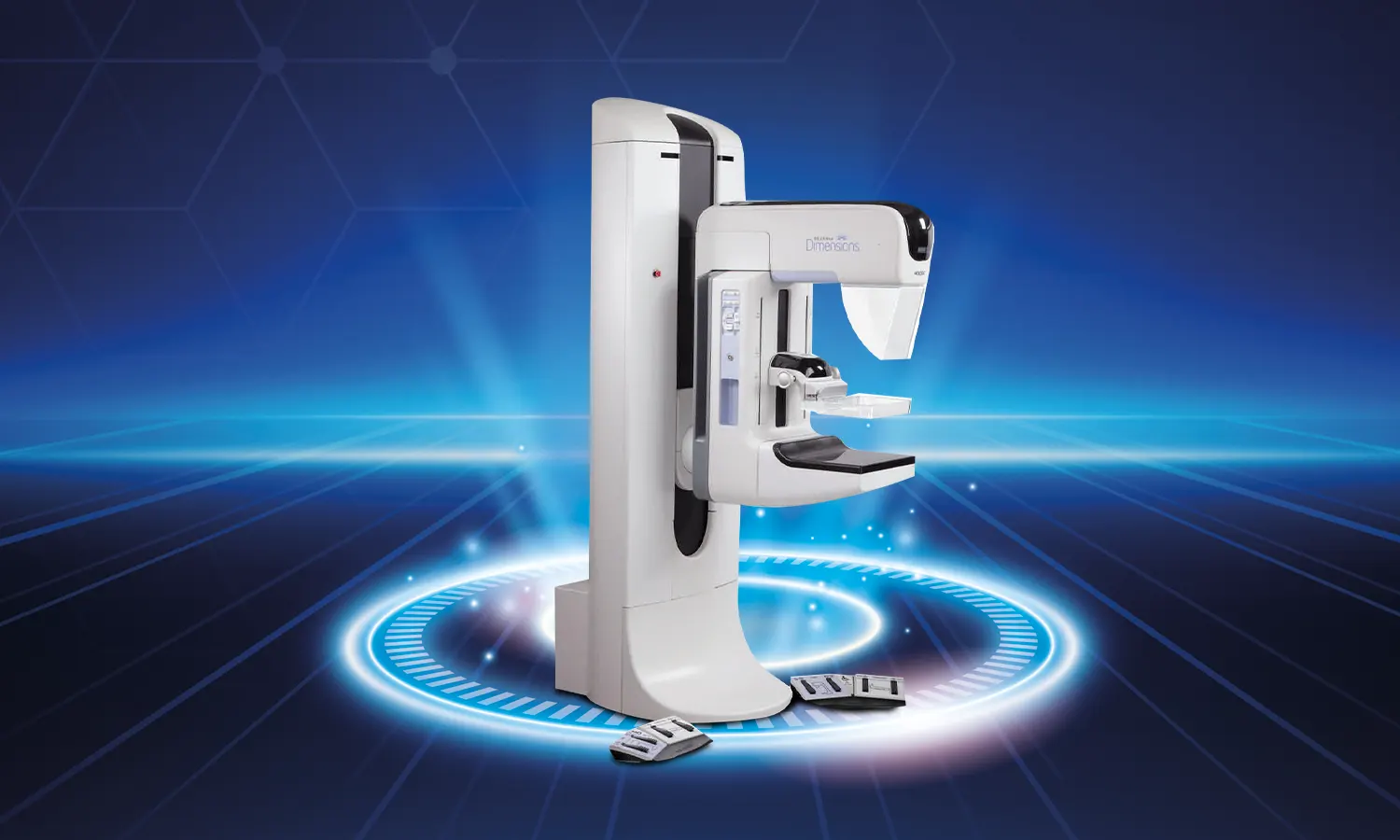
Breast cancer remains one of the most prevalent forms of cancer affecting women worldwide, emphasizing the critical importance of effective screening methods. Among the various techniques employed for breast cancer detection, mammography stands out as the cornerstone of early diagnosis. Over the years, mammography has evolved, with advancements in technology continually enhancing its capabilities. One such advancement is 3D Breast Tomosynthesis, a groundbreaking technique that revolutionizes the way breast tissue is imaged and analyzed.
Unlike traditional mammography, which captures two-dimensional images of the breast, 3D Breast Tomosynthesis offers a three-dimensional perspective, providing radiologists with a more comprehensive view of breast tissue architecture. This innovative approach involves acquiring multiple thin slices or sections of the breast from different angles. These slices are then reconstructed into a three-dimensional image dataset, allowing for a detailed examination of breast tissue structures.
The digital nature of 3D Breast Tomosynthesis facilitates precise localization and characterization of abnormalities within the breast. By examining breast tissue slice by slice, radiologists can identify subtle lesions that may be obscured or difficult to detect on conventional mammograms. This level of detail is particularly advantageous in distinguishing between benign and malignant lesions, reducing the incidence of false positives and unnecessary biopsies.
Numerous clinical studies have demonstrated the clinical utility of 3D Breast Tomosynthesis in improving breast cancer detection rates and patient outcomes. One of the largest studies conducted on this subject, involving 33,000 patients over a three-year period, yielded compelling results. Researchers found that lesions detected using 3D Breast Tomosynthesis were characterized more easily, resulting in fewer recalls for additional imaging. Moreover, the technique led to an increase in the detection of invasive cancers, particularly small, early-stage tumors that may have been overlooked with conventional mammography.
In addition to enhancing cancer detection, 3D Breast Tomosynthesis offers other significant benefits. By reducing false positives and unnecessary recalls, it helps alleviate patient anxiety and streamlines the diagnostic process. Furthermore, the technique enables more accurate assessment of lesions identified during screening, leading to improved decision-making regarding further evaluation and treatment.
Despite its advanced capabilities, concerns regarding radiation exposure associated with 3D Breast Tomosynthesis have been addressed through technological innovations. Modern devices utilize low-dose imaging protocols, ensuring that radiation exposure remains within safe limits comparable to standard mammography.
In conclusion, 3D Breast Tomosynthesis represents a major advancement in breast cancer screening technology, offering improved detection rates, enhanced diagnostic accuracy, and better patient outcomes. As research continues to validate its efficacy and safety, this innovative technique holds promise for transforming the landscape of breast cancer diagnosis and management.
© 2026 Copyright by Private Koru Hospital. All rights reserved.