- Call Today +90 312 911 77 77
- Open Hour 09:00 AM to 18:00 PM
Micro Ultrasound Fusion Biopsy represents an advanced diagnostic method that merges two imaging technologies, micro-ultrasound, and MRI, to pinpoint and assess prostate cancer lesions for biopsy. This approach marks a significant advancement over conventional transrectal ultrasound (TRUS) biopsies, which might overlook small or challenging-to-detect tumors, leading to potential overdiagnosis or overtreatment of prostate cancer.
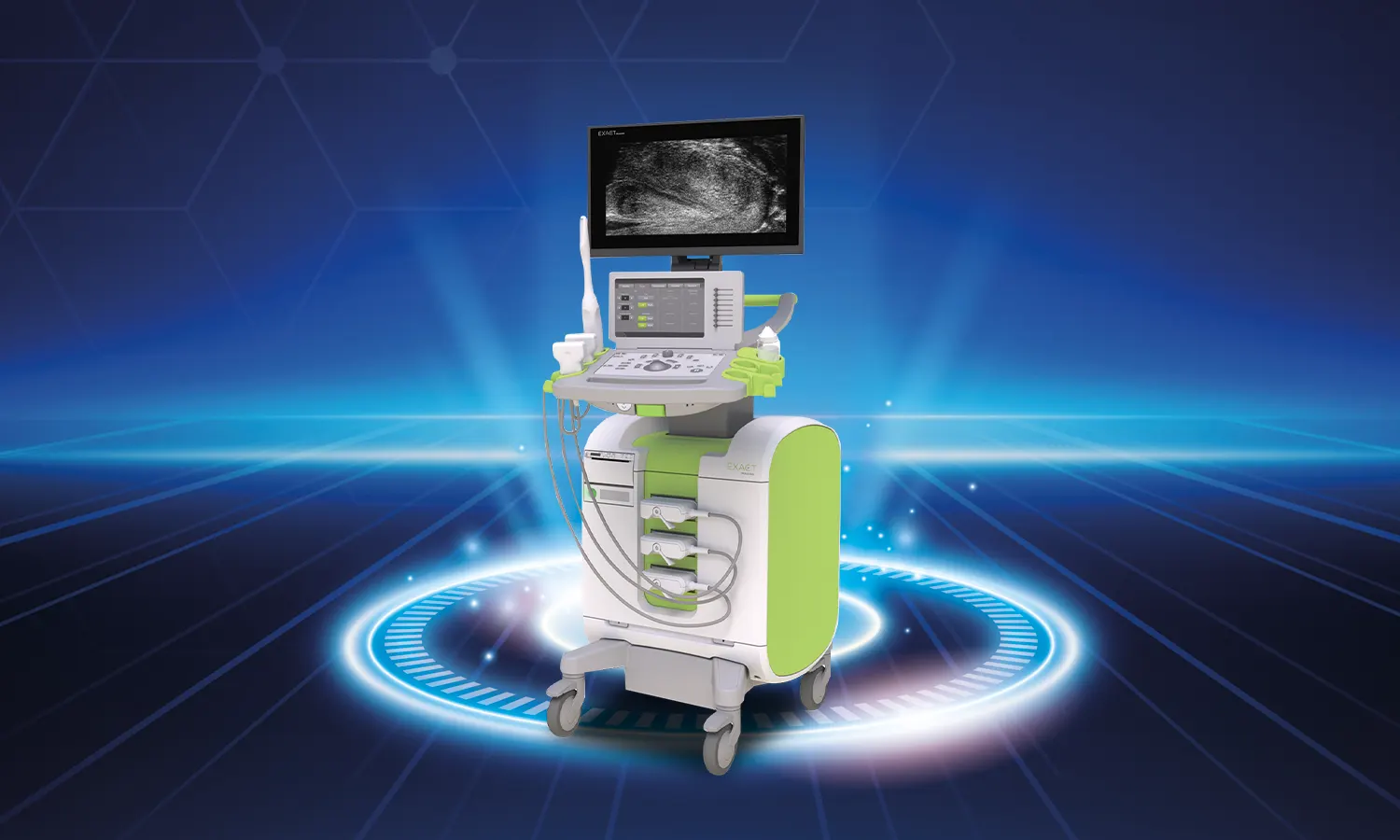
In the micro-ultrasound fusion biopsy, a urologist utilizes a micro-ultrasound probe to generate real-time, high-resolution images of the prostate gland. These images are then fused with previously acquired MRI images to provide a more detailed perspective of the gland and any suspicious lesions. The fused images guide the biopsy needle accurately to the tumor location, enhancing biopsy precision and minimizing the risk of complications.
Although Micro Ultrasound Fusion Biopsy is a relatively recent technology with limited adoption, it exhibits promising outcomes in detecting prostate cancer, particularly in cases involving elevated prostate-specific antigen (PSA) levels or previous negative biopsies. It's essential to acknowledge that, while this biopsy technique enhances accuracy, it is not flawless, and false negatives or positives can still occur.
The primary distinction between Micro Ultrasound Fusion Biopsy and traditional methods like transrectal ultrasound (TRUS) biopsy lies in the utilization of multiple imaging modalities to precisely target suspicious areas within the prostate gland.
In TRUS biopsy, a transrectal ultrasound probe generates prostate images, and a biopsy needle collects tissue samples through the rectum. However, TRUS biopsy has limitations in detecting and characterizing suspicious lesions, often resulting in inaccuracies. Micro Ultrasound Fusion Biopsy utilizes micro-ultrasound and MRI to provide a more accurate view, reducing the likelihood of false results.
Another key difference is that Micro Ultrasound Fusion Biopsy is typically performed under local anesthesia, without the need for antibiotics or sedatives, making it less invasive and more tolerable for patients.
Both Micro Ultrasound Fusion Biopsy and Magnetic Resonance Imaging (MRI) offer distinct advantages in prostate cancer detection and diagnosis:
In summary, combining Micro Ultrasound Fusion Biopsy with MRI provides a comprehensive approach to prostate cancer detection and diagnosis.
Micro Ultrasound (micro-US) is a high-resolution technique without specific stages, but it involves several steps:
Micro Ultrasound is a minimally invasive, accurate technique for imaging and biopsy.
The success rate of Micro Ultrasound (micro-US) varies based on the case and the procedure's purpose. It excels in detecting small tumors or suspicious areas within the prostate gland, particularly when combined with targeted biopsy techniques like Micro Ultrasound Fusion Biopsy or cognitive fusion biopsy. Success rates can reach up to 90%.
The expertise of the urologist or radiologist performing the procedure also influences the success rate. Individual factors, such as age, overall health, and tumor characteristics, contribute to the overall success in detecting prostate cancer.
In summary, Micro Ultrasound is highly accurate and minimally invasive, providing valuable insights into prostate cancer detection.
Micro Ultrasound Fusion Biopsy, combining micro-ultrasound and MRI, offers several advantages over traditional biopsy methods:
Micro Ultrasound Fusion Biopsy is a valuable tool in prostate cancer detection and diagnosis, offering increased accuracy and a more patient-friendly experience.
Micro Ultrasound Fusion Biopsy is a minimally invasive procedure combining micro-ultrasound with MRI for targeted prostate biopsies:
Micro Ultrasound Fusion Biopsy combines high-resolution images with detailed MRI information, providing a comprehensive view for accurate prostate cancer diagnosis and staging.
© 2024 Copyright by Private Koru Hospital. All rights reserved.