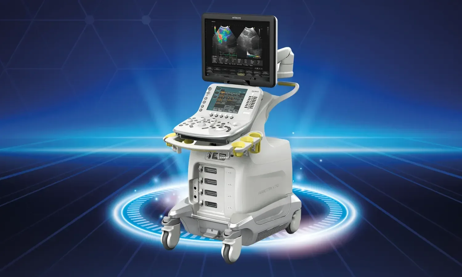
Advanced ultrasonography is an enhanced imaging method that allows for a detailed evaluation of the fetus during pregnancy. By using high-resolution ultrasonography devices, fetal structures are examined in greater detail, and potential abnormalities are detected. This method provides more detailed images than standard pregnancy ultrasonography and allows for the assessment of various parameters such as fetal development, amniotic fluid volume, and placental function. Fetal therapy, on the other hand, is a set of advanced medical practices that enable intervention for detected anomalies during the prenatal period. These developments in the field of perinatology have become indispensable tools for protecting the health of both mother and baby in high-risk pregnancies.
Working Principle of Advanced Ultrasonography and Fetal Therapy
Advanced ultrasonography is based on the integrated use of sophisticated techniques such as three-dimensional (3D) and four-dimensional (4D) imaging, color Doppler, and fetal echocardiography. Through high-frequency sound waves, fetal organs can be examined at a millimetric level. Thanks to Doppler technology, blood flow between the fetus and the placenta can be evaluated. In fetal therapy aimed at addressing detected issues, direct intervention is performed on the baby by entering the uterus. These treatments are carried out under ultrasound guidance, making it possible to perform life-saving procedures before the baby is born. With a method called fetoscopy, a camera can be inserted into the uterus to perform direct visual intervention.
In Which Situations Is It Used?
Detection of Fetal Anomalies
Many structural anomalies such as congenital heart diseases, neural tube defects, kidney and bladder anomalies, and skeletal system disorders can be detected with advanced ultrasonography. Early diagnosis ensures that birth planning is carried out in appropriate centers and that prenatal treatment is initiated when necessary. Detailed screenings performed during the first and second trimesters play an important role in the detection of chromosomal anomalies.
Conditions Requiring Fetal Surgery
In cases such as spina bifida, congenital diaphragmatic hernia, lower urinary tract obstruction, and twin-to-twin transfusion syndrome, prenatal surgical intervention can be life-saving. These procedures are performed by experienced teams in specialized centers. Thanks to fetal surgery, the postnatal prognosis of anomalies corrected in the womb is significantly better.
Fetal Anemia and Blood Diseases
Fetal anemia may develop due to reasons such as Rh incompatibility or parvovirus infection. In this case, the baby is given blood via intrauterine transfusion. Fetal anemia can be diagnosed with Doppler ultrasound, and the response to treatment can be monitored. Middle cerebral artery Doppler measurements are considered the gold standard method for diagnosing fetal anemia.
High-Risk Pregnancy Follow-up
Chronic maternal diseases such as diabetes, hypertension, and autoimmune diseases, or multiple pregnancies, are closely monitored with advanced ultrasonography. Problems such as placental insufficiency and intrauterine growth restriction can be detected at an early stage. This allows for the decision of delivery at the appropriate time and prevents adverse outcomes for both the mother and the baby.
Advantages of Advanced Ultrasonography and Fetal Therapy
- Detailed fetal assessment: Fetal organs and structures can be examined at a millimetric level thanks to high-resolution images.
- Early anomaly detection: Potential fetal anomalies can be detected at an early stage, and necessary precautions can be taken.
- Prenatal treatment opportunity: It is possible to treat some diseases before the baby is born.
- Information for the family: The family receives detailed information about the baby's condition and prepares for the birth process.
- Guidance in birth planning: The appropriate place, time, and method of delivery can be determined.
- Increase in quality of life: With early intervention, the baby's postnatal quality of life can be significantly improved.
- Non-invasive examination: It is a safe and painless imaging method for both mother and baby.
- Repeatability: It can be applied as often as desired throughout the pregnancy.
Fetal Therapy Methods
Fetal surgery consists of surgical procedures performed on the fetus during the prenatal period and is used in the treatment of certain anomalies. During medical treatment, drug therapies directed at the fetus can be administered throughout the pregnancy. Among intrauterine interventions, procedures such as amniocentesis and cordocentesis are used to obtain information about the fetus's condition and perform necessary interventions. Fetal transfusions allow for blood transfusion to the fetus to treat certain blood diseases. Laser photocoagulation is a method successfully applied in the treatment of transfusion syndrome in twin pregnancies. With shunt placement procedures, fetal bladder or lung fluid accumulations are drained to ensure organ development.
Treatment Process
The process begins with routine ultrasound screenings performed in the early stages of pregnancy. When a suspected condition is detected, a referral is made for advanced ultrasonography. As a result of the detailed examination, additional tests such as genetic tests, fetal MRI, or echocardiography may be requested if necessary. An appropriate treatment plan is determined for cases discussed in a multidisciplinary council. The council includes a perinatologist, pediatric surgeon, pediatric cardiologist, and geneticist. In cases requiring fetal therapy, the procedure is usually performed under mild sedation with ultrasound guidance. After treatment, the condition of the mother and baby is closely monitored. Delivery is planned at a tertiary center where an experienced team is available.
Points to Consider
Fetal therapy may not be suitable for every pregnancy, and the condition of the mother and baby should be evaluated together when making a decision. Procedures involve certain risks such as miscarriage or preterm birth, and the family must be provided with detailed information. Regular check-ups should be attended, and recommended tests should not be skipped. Receiving genetic counseling and providing psychological support to the family are of great importance throughout the process. Smoking, alcohol, and the use of medications not approved by a doctor should be avoided during pregnancy. The use of folic acid supplementation and recommended vitamin supplements is effective in preventing neural tube defects. Participation in prenatal classes enables the family to approach the process more consciously.
Three- and Four-Dimensional Imaging
Three-dimensional ultrasound imaging allows for a detailed examination of the fetal face, hands, feet, and external genital organs. Facial anomalies such as cleft lip and palate can be detected much more easily with this method. Four-dimensional ultrasound allows for the observation of fetal movements in real-time. In this way, fetal behavior assessment can be performed, and families can build a stronger emotional bond with their babies. With a technology called STIC, all parts of the fetal heart are obtained in 3D in a single scan, allowing for detailed analysis. The detection of congenital heart diseases has been significantly facilitated by this.
As Koru Hospital, we provide the highest level of service in the follow-up of high-risk pregnancies and fetal therapy with our perinatology specialists and state-of-the-art ultrasound devices. We continue to plan the safest and healthiest birth process together for our expectant mothers and their babies.