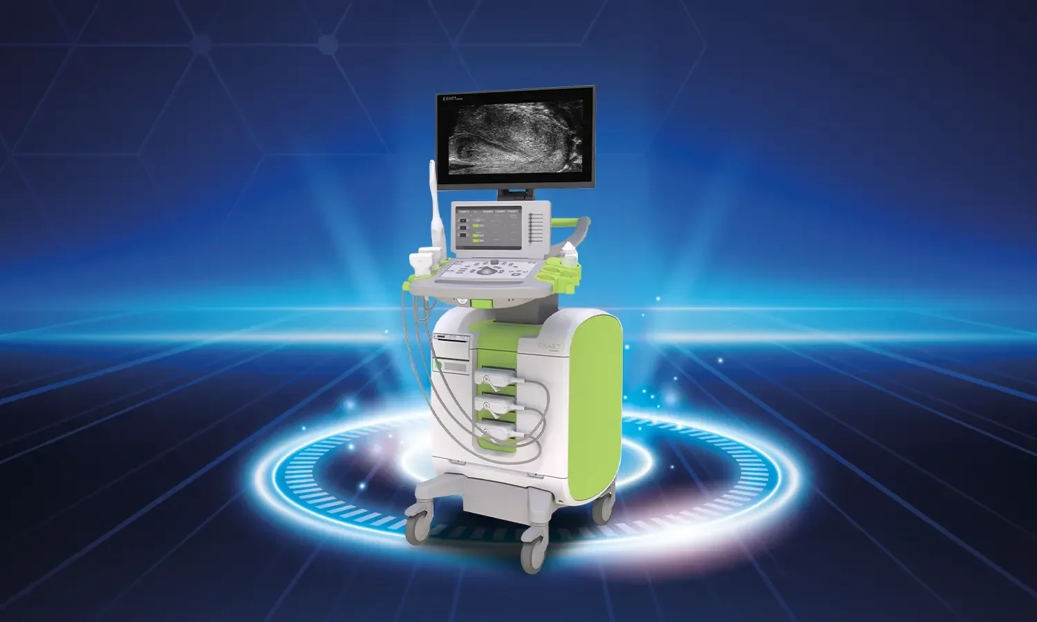
Endoscopic ultrasonography (EUS) is an advanced medical imaging method used for the detailed visualization and examination of the digestive system and surrounding tissues. This method combines endoscopy and ultrasonography, making it possible to obtain high-resolution images of internal organs and tissues. By overcoming the limitations of traditional endoscopy, which only visualizes the superficial mucosa, EUS allows for the evaluation of intramural structures and adjacent organs, becoming an indispensable diagnostic tool in modern gastroenterology.
How Does EUS Work?
Endoscopic ultrasonography involves inserting an endoscope, a thin and flexible tube, into the body through the mouth or the anal region (rectum). An ultrasound probe is located at the tip of the endoscope; this probe is used to image organs and tissues inside the body using sound waves. Ultrasound waves reflect off body tissues to create a detailed image of the organs. The probe's ability to work in extremely close proximity to the target organ allows for the differentiation of muscle layers and the examination of deep structures that superficial ultrasound cannot evaluate. During EUS, procedures such as fine-needle aspiration (FNA) and fine-needle biopsy (FNB) can also be performed, allowing for tissue sampling from suspicious areas.
The EUS system can clearly distinguish the five separate layers of the digestive tract wall; this plays a critical role in treatment planning, especially in cancer staging, by determining how far a tumor has progressed into the layers. Furthermore, it is among the most sensitive methods for providing a detailed evaluation of organs that are difficult to reach via the skin, such as the pancreas.
In Which Diseases Is It Used?
- Diagnosis and staging of tumors: The size, depth, and spread of tumors in digestive system organs such as the stomach, esophagus, pancreas, and rectum are evaluated.
- Pancreatic cysts and masses: It plays a critical role in distinguishing whether pancreatic lesions are benign or malignant.
- Lymph node biopsy: Fine-needle biopsy can be taken from lymph nodes to determine cancer spread and for staging.
- Biliary tract diseases: Used to detect stones, strictures, and tumors in the bile ducts.
- Submucosal lesions: Examination of hidden masses (such as GIST) within the stomach or intestinal wall.
- Differential diagnosis of chronic pancreatitis and pancreatic cancer
- Mediastinal lymph node evaluation: An important tool in lung cancer staging
- Evaluation of anorectal diseases
- EUS-guided interventional procedures: Pancreatic pseudocyst drainage, celiac plexus block, biliary drainage
Advantages of EUS
- High-resolution and detailed imaging
- Ability to evaluate all layers of the digestive tract wall in detail
- Opportunity for both diagnosis and tissue sampling in the same session
- Minimally invasive approach compared to surgical biopsy
- Evaluation of surrounding organs and vascular structures
- Detection of early-stage tumors and accurate staging
- Provision of detailed information that guides treatment planning
- Being one of the most sensitive imaging methods for pancreatic diseases
- Guidance for therapeutic procedures without the need for surgery
- Safe imaging method that does not involve radiation
Comparison with Other Imaging Methods
Methods such as abdominal ultrasound, CT, and MRI are also used to evaluate the digestive system and surrounding organs; however, EUS offers superior resolution, especially in the evaluation of the pancreas and the digestive tract wall, due to its ability to work in extremely close proximity to the target organ. The limitations of CT (radiation exposure) and MRI (high cost and contraindications) do not apply to EUS. At the same time, EUS is the preferred first-line modality in evidence-based guidelines for the evaluation of small pancreatic lesions and submucosal tumors. In addition to these, EUS-guided fine-needle aspiration offers the possibility of a definitive pathological diagnosis without the need for surgical biopsy, saving time in treatment planning.
Treatment and Procedure Process
Before the EUS procedure, the patient is required to fast for a certain period; this ensures that the stomach and intestines are empty and that image quality is high. At least 6 hours of fasting is required for upper system EUS, and bowel preparation is required for lower system EUS. The procedure is usually performed under sedation; the patient is not awake and does not feel pain or discomfort. The endoscope is inserted orally or anally, and once the target area is reached, the ultrasound probe is activated to image the tissues in detail. When deemed necessary, samples are taken from suspicious areas with a fine needle under real-time ultrasound guidance; the samples taken are sent for pathological examination in the same session. The procedure duration is usually between 30-60 minutes.
After the effects of sedation wear off, the patient is discharged; they can gradually return to their daily activities within the same day. Pathology results are usually ready within a few business days, and the results guide the treatment plan. Evaluating the case together with surgical, oncology, and radiology teams through a multidisciplinary approach is important for determining the most appropriate treatment option.
Points to Consider
Although endoscopic ultrasonography is an invasive procedure, it is generally safe for the patient, and serious complications are rare. However, temporary side effects such as mild sore throat, bloating, or gas may occur after the procedure. Very rarely, risks such as bleeding, perforation, acute pancreatitis, or infection may occur. Medications used before the procedure (especially blood thinners), bleeding disorders, heart or lung diseases, and allergies must be reported to the physician. Due to sedation, patients should not drive, make important decisions, or should be accompanied by an escort on the day of the procedure. Since the success of EUS is closely related to the experience of the practitioner, it is of great importance that it is performed by experienced endoscopists in well-equipped centers.
EUS at Koru Hospital
As Koru Hospital, we provide high-accuracy services in the diagnosis of digestive system and surrounding organ diseases with our expert team experienced in gastroenterology and interventional endoscopy and our advanced technology EUS equipment. We perform EUS applications for our patients accompanied by safe sedation conditions, our modern endoscopy unit, and the fast and reliable support of our pathology laboratory. Each patient's clinical history is meticulously evaluated, the diagnostic process is managed with the participation of surgical, oncology, and radiology teams through a multidisciplinary approach, and the treatment plan is coordinated by the same team when necessary. We care about being with our patients at every stage, from before the procedure to the pathology results, and facilitating the process with accurate information and psychological support. As Koru Hospital, our goal is to increase treatment success by detecting all related diseases, especially those of the digestive system, at an early stage and to offer our patients a safe and comfortable diagnostic process.