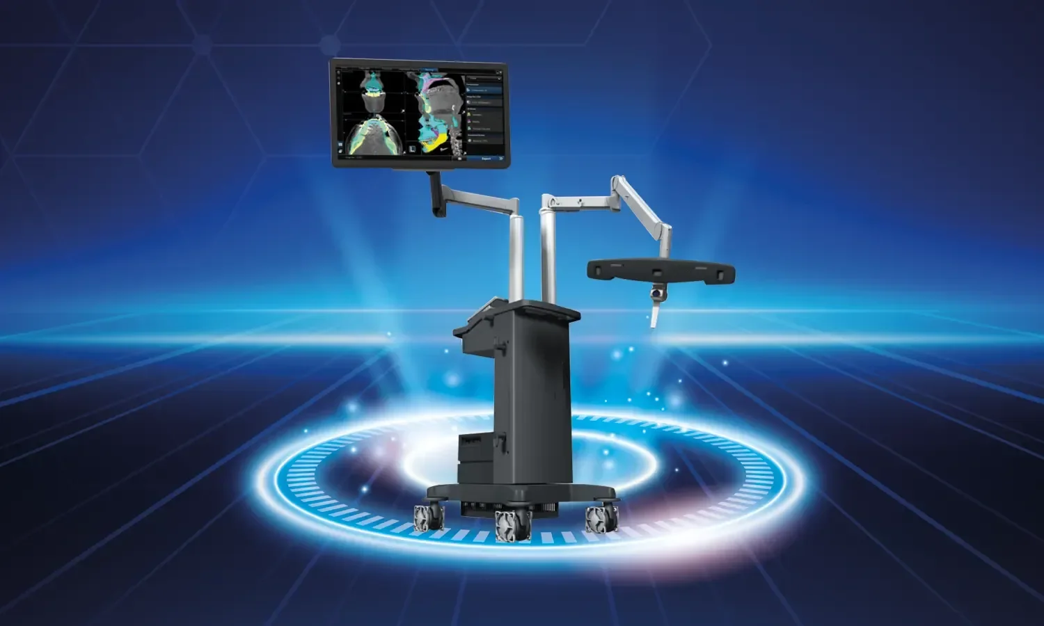
Early and accurate diagnosis of vascular diseases is the most important factor directly affecting treatment success. While traditional angiography methods show the vascular structure only in two dimensions, the 4D angiography technology developed in recent years can display vessels both in three dimensions and with blood flow dynamics over time. In this way, clinicians can evaluate not only the anatomy of the disease but also its functional behavior. The method has brought revolutionary innovations, especially in the treatment planning of complex vascular anomalies.
What is 4D Angiography?
4D (four-dimensional) angiography is an advanced vascular imaging method obtained by adding a time component to three-dimensional imaging. While a single moment is displayed in classical angiography, the filling and emptying processes of the vessel are tracked in small fractions of a second in 4D angiography. Thus, the direction, speed, turbulence, and circulation timing of blood flow can be clearly measured. This feature provides a unique advantage in the treatment planning of complex vascular anomalies and gives the physician information that static images cannot offer.
Working Principle
The procedure is performed with high-speed digital subtraction angiography devices. The flow of the contrast agent injected into the vessel via a catheter is imaged at a speed of dozens of frames per second with multi-directional detectors. The obtained data is processed with computer software and converted into a moving three-dimensional model. The physician can examine this model by rotating it from every angle and evaluate the details by zooming into the desired area. Thanks to the color mapping of flow direction and speed, abnormal flow patterns are detected within seconds.
In Which Diseases Is It Used?
4D angiography plays a decisive role in the diagnosis and treatment planning of many complex vascular diseases:
- Brain aneurysms: Evaluation of the aneurysm neck and filling pattern
- Arteriovenous malformations: Differentiation of feeding and draining vessels
- Dural arteriovenous fistulas: Detection of abnormal shunt points
- Carotid and vertebral artery stenoses: Measurement of flow dynamics
- Aortic diseases: Monitoring of aneurysms and dissections
- Collateral circulation assessment: Mapping of bypass routes for occluded vessels
- Liver and kidney vascular pathologies
- Pre-surgical and interventional mapping
- Pulmonary embolism and pulmonary vascular diseases
- Evaluation of vascular tumors
Advantages of 4D Angiography
Compared to classical 2D and 3D methods, 4D angiography offers many unique advantages:
- Temporal resolution: Flow dynamics in the vessel are shown in fractions of a second.
- Superior anatomical detail: Vascular structures can be examined from every angle.
- Accurate treatment planning: A roadmap is created before surgical or endovascular treatment.
- Low contrast agent requirement: Comprehensive information is obtained in a single session.
- Clarity in complex cases: Intertwined vascular structures can be separated.
- Treatment follow-up: Post-procedural flow changes are monitored objectively.
- Multi-disciplinary harmony: Surgeons, radiologists, and neurologists share the same data.
- Single procedure for the patient: Diagnosis and, if necessary, treatment can be performed in the same session.
- High success rate: Visual accuracy supports physician decision-making.
How Does the Examination Process Proceed?
Before the procedure, the patient's detailed history is taken. Routine tests such as kidney function tests, blood tests, and ECG are completed. A history of contrast agent allergy and medications used are questioned. A 4-6 hour fasting period is required before the procedure. Blood-thinning medications used must be reported to the physician; these medications are temporarily discontinued if necessary.
Procedure Phase
4D angiography is performed in the angiography unit. Under local anesthesia, a catheter is usually inserted through the femoral artery and advanced to the target vessel. High-speed imaging is initiated while the contrast agent is administered. Images are processed instantly on a computer. The procedure takes an average of 30-60 minutes; it is shorter when performed for diagnostic purposes. If treatment is required, endovascular intervention can be performed in the same session.
Post-Procedure Process
Pressure is applied to the vascular access site. The patient is discharged after a few hours of bed rest. The results are evaluated by experienced radiologists and relevant branch physicians to create a treatment plan. After the patient returns home, checking the access site and using the recommended medications are important.
Points to Consider
Some important points should be considered before and after the procedure:
- A history of contrast agent allergy must be reported to the physician.
- Additional precautions are taken for individuals with kidney disease.
- In diabetic patients, metformin-type medications may be temporarily discontinued.
- Consuming plenty of fluids after the procedure accelerates contrast excretion.
- Heavy activity should be avoided at the vascular access site for 24 hours.
- Any suspicion of pregnancy must be reported in advance.
- Blood thinners used must be managed with physician approval.
- A physician should be consulted in case of swelling, increased heat, or bruising at the access site.
- Follow-up imaging should be performed at the planned times.
Revolutionary Advantages of Treatment Planning with 4D Angiography
Beyond being just an imaging method, 4D angiography offers the physician a detailed simulation opportunity before treatment. Especially in brain AVM treatments, the sequence in which feeding vessels will be embolized and which microcatheter will be used at what angle can be planned in advance on these images. Thus, the actual intervention is performed faster, safer, and more successfully. In aortic pathologies, stent graft selection and embolic protection device planning in carotid stenting are also optimized with 4D data.
Comparative Imaging and Follow-up
4D angiography is also of great value in post-treatment follow-up. How an embolized AVM shows a flow pattern over time, whether there is residual filling after aneurysm treatment, and the long-term performance of a carotid stent can be objectively evaluated with 4D data. Thanks to comparative analysis, anatomical changes that develop after treatment are detected in the early period, and additional intervention is planned if necessary. This approach provides long-term safety for patients and prevents unexpected complications.
As Koru Hospital, we offer a unique service in the diagnosis and treatment of complex vascular diseases with our advanced technological infrastructure in the field of 4D angiography and the international experience of our interventional radiology team. Thanks to our multidisciplinary approach, we determine the most accurate treatment strategy by evaluating each patient's individual condition in our council consisting of neurology, neurosurgery, cardiovascular surgery, and radiology specialists. Thanks to our hybrid operating room infrastructure, diagnosis and treatment can be applied in the same session when necessary; thus, we provide our patients with time and comfort advantages. We consider it our duty to protect your health by using the opportunities offered by modern medicine at the highest level and to continue serving as a leading center in the early diagnosis and effective treatment of vascular diseases.