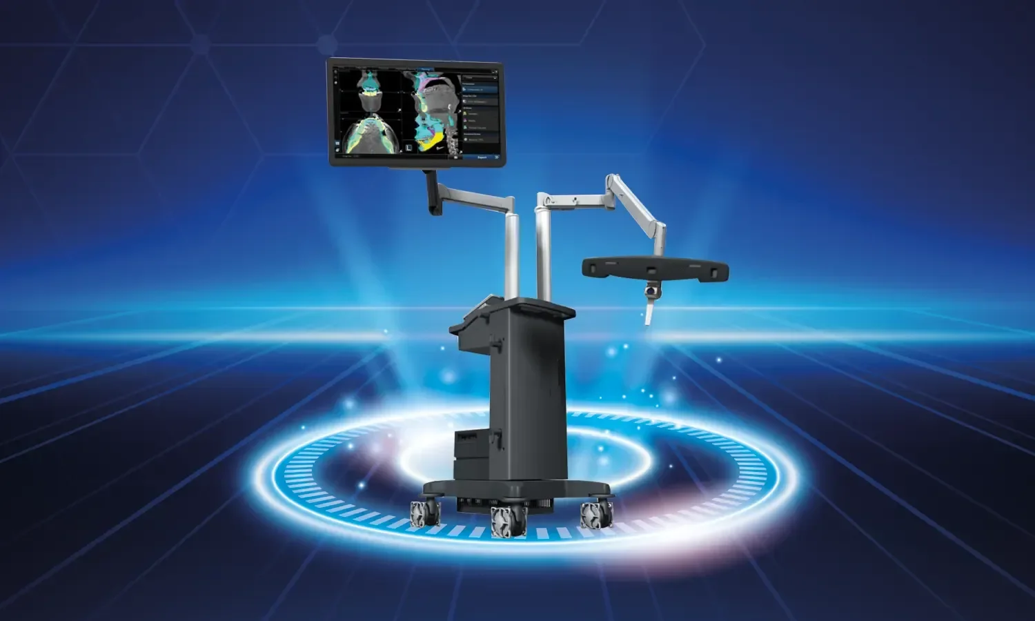
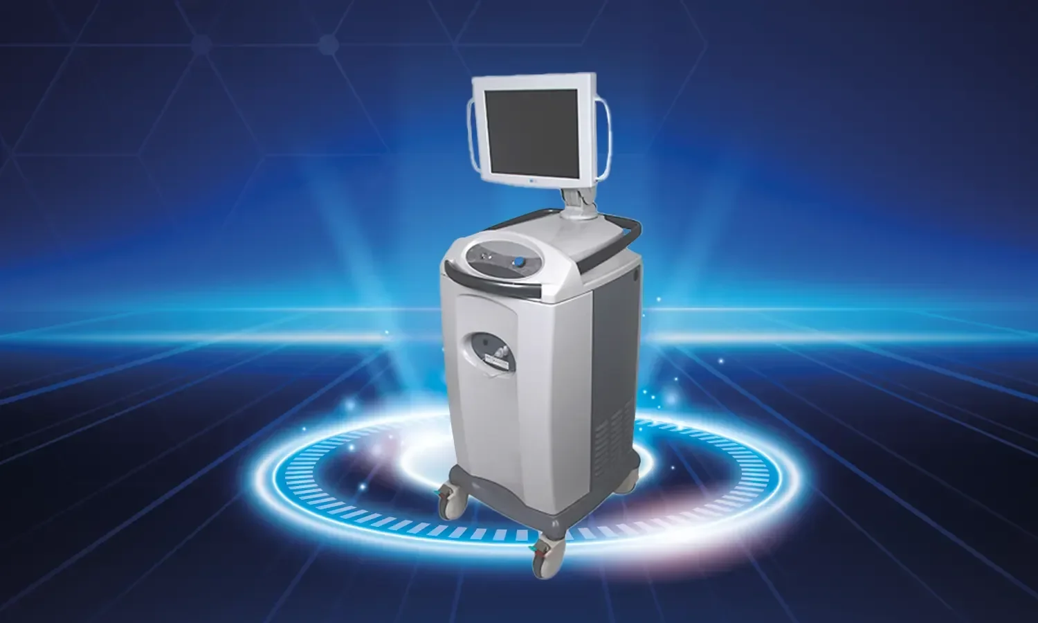
In neurosurgery, the surgical microscope is an indispensable tool that allows surgeons to obtain a clearer and more detailed view during operations. Playing a significant role in the development of modern neurosurgery, surgical microscopes offer the surgeon high magnification, superior illumination, and ergonomic working conditions during brain and spinal cord surgeries that require millimetric precision. As a result, the success rate of interventions performed in complex anatomical regions is significantly increased.
What is a Surgical Microscope and How Does It Work?
A surgical microscope is a high-tech device equipped with optical systems and specialized light sources that visualize the surgical field with a high magnification ratio. Thanks to its dual-eyepiece structure, the device provides three-dimensional depth perception, allowing the surgeon to navigate safely between delicate tissues. Modern surgical microscopes can be used in conjunction with fluorescence imaging, intraoperative angiography (ICG), integrated neuromonitoring, and image recording systems. These auxiliary technologies facilitate the differentiation of cancerous tissue or vascular structures from healthy tissue during surgery.
The optical system of the surgical microscope offers the ability to switch between different magnification ratios; the surgeon can fluidly transition between a general view and a detailed close-up as needed. With LED or xenon light sources, the surgical field is illuminated with a quality similar to daylight. Furthermore, thanks to assistant eyepieces, other members of the team can simultaneously follow the same view as the surgeon; this enhances both team coordination and training opportunities.
Functions of the Surgical Microscope
- High magnification ratio: Shows the surgical field with a very high magnification ratio, ensuring small structures and details are clearly visible.
- Differentiating capability: Makes it easier to distinguish healthy tissues from lesions and abnormal structures.
- Superior illumination: Even narrow and deep areas can be illuminated with specially designed lighting systems.
- Ergonomic design: Allows surgeons to work comfortably during long-duration surgeries.
- Fluorescence imaging: Techniques such as ICG angiography and 5-ALA allow for real-time visualization of vascular permeability or tumor tissue.
- Image recording and training support: Recording the surgery provides opportunities for training and evaluation.
- Three-dimensional depth perception: The dual-eyepiece structure allows for clear perception of the distance between tissues.
- Assistant and team view sharing: Simultaneous viewing improves team coordination.
- Neuronavigation integration: Real-time tracking of the preoperatively planned route.
In Which Surgeries Is It Used?
- Brain tumor surgeries (glioma, meningioma, acoustic neuroma, pituitary adenoma)
- Aneurysm and arteriovenous malformation (AVM) surgery
- Spine and spinal cord tumor surgery
- Microdiscectomy and disc surgery
- Peripheral nerve surgery and nerve repairs
- Microsurgical repairs in plastic surgery
- Middle ear and inner ear surgeries in otorhinolaryngology (ENT)
- Eye surgery (ophthalmology) procedures
- Hand and microsurgery applications
- Vascular repairs and microvascular anastomoses
- Skull base surgery
- Pediatric neurosurgery
Advantages of the Surgical Microscope
- Superior success in preserving delicate anatomical structures
- Three-dimensional imaging enabling the application of microsurgical techniques
- Less tissue trauma and lower risk of complications
- Clearer determination of tumor margins
- Reduced risk of ischemia by preserving vascular structures
- Efficient use of surgical time
- Reduced surgeon fatigue and increased focus
- Improvement of postoperative functional outcomes
- Higher rate of completeness in tumor resection
- Increased long-term quality of life through the preservation of nerve and vascular structures
- Significant role in assistant training
Treatment Process and Surgical Approach
Prior to operations performed using a surgical microscope, the anatomical relationship of the lesion is evaluated through detailed imaging studies (MRI, CT, angiography). When necessary, three-dimensional surgical planning is performed by combining this with neuronavigation systems and intraoperative imaging. The patient's detailed neurological examination, medications used, and accompanying diseases are reviewed. Following an anesthesia consultation, the appropriate surgical approach is determined through a multidisciplinary evaluation.
During surgery, the procedure is performed while protecting small vascular and nerve structures thanks to the magnification and illumination features of the microscope. The additional technologies used (ICG angiography, fluorescence-guided resection, intraoperative neuromonitoring) increase the safety and success of the surgery. Especially in brain tumor surgeries, 5-ALA fluorescence allows for more complete resection by distinguishing tumor cells from healthy tissue. In microvascular anastomoses, vascular permeability is verified in real-time with ICG angiography. Postoperative follow-ups are planned according to the type of lesion and the intervention performed; the neurological status is closely monitored, and physical therapy and rehabilitation processes are initiated when necessary.
Points to Consider
The success of surgeries performed with a surgical microscope is closely related to the surgeon's experience, team coordination, the patient's anatomical structure, and the characteristics of the lesion. Although modern devices provide great advantages, patient selection, surgical planning, and the postoperative care process are equally important. Completing recommended tests during the preoperative preparation process, informing the physician about medications used, and strictly adhering to postoperative follow-up checks directly affect the quality of the recovery process. Obtaining a clearer and more detailed view helps surgeons move more accurately and reduces the risk of complications. Changes in neurological status during the postoperative period should be closely monitored, and adherence to recommended medication treatments and physical therapy programs should be ensured. The recovery process after neurosurgery can be long; patience and regular follow-up are the cornerstones of successful results.
Surgical Microscope at Koru Hospital
As Koru Hospital, we provide services with our high-tech surgical microscopes and experienced surgical staff in all branches requiring microsurgery, primarily neurosurgery. In our modernly equipped operating rooms, we provide our patients with surgical opportunities at high safety standards by integrating supportive technologies such as neuronavigation, intraoperative imaging, fluorescence-guided resection, and neuromonitoring with the surgical microscope. We continue to stand by our patients with our multidisciplinary approach and our follow-up process that lasts from before surgery until discharge. Our expert staff in neurosurgery, orthopedics, otorhinolaryngology, ophthalmology, plastic surgery, and other related branches work together to provide our patients with the highest level of medical service. We manage the postoperative recovery process holistically with our modern intensive care units, experienced rehabilitation team, and psychological support services. As Koru Hospital, our goal is to increase surgical success and protect our patients' quality of life at the highest level by using the microsurgical opportunities offered by modern medicine in the most accurate way.