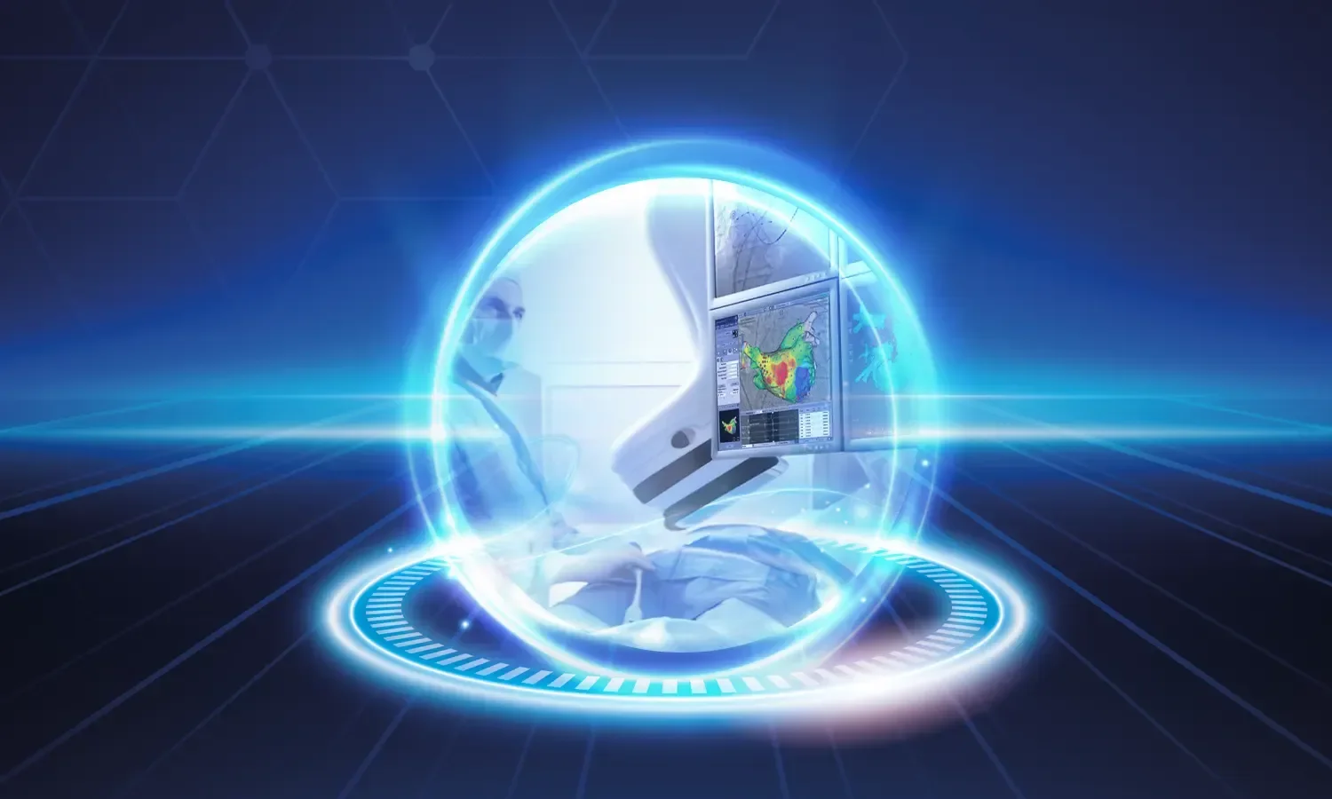
C-Arm skopi, gerçek zamanlı röntgen görüntüleme yapabilen, C harfi şeklindeki taşıyıcı kola sahip mobil radyoloji cihazıdır. Gastroenteroloji başta olmak üzere ortopedi, kardiyoloji, üroloji ve girişimsel radyoloji gibi pek çok branşta vazgeçilmez bir araç olan bu sistem, hekime ameliyat veya işlem sırasında anlık görüntü sağlayarak girişimlerin güvenli ve doğru şekilde yapılmasını mümkün kılar. Özellikle endoskopik retrograd kolanjiopankreatografi (ERCP) gibi karmaşık girişimsel işlemlerde altın standart görüntüleme yöntemidir. Açık cerrahiye alternatif minimal invaziv yaklaşımlara olanak tanıyan C-Arm skopi, modern tıpta hasta konforu ve tedavi başarısının artmasına önemli katkı sağlamaktadır.
C-Arm Skopi Nedir ve Nasıl Çalışır?
C-Arm skopi, X-ışını kaynağı ve görüntü dedektörü içeren bir C-kollu yapı, yüksek çözünürlüklü monitörler ve kontrol konsolundan oluşur. Cihazın C şeklindeki tasarımı, hastanın çevresinde 360 derece dönüş yapabilmesini sağlar ve böylece istenen anatomik açıdan görüntü alınabilir. Modern C-Arm sistemleri dijital görüntü işleme, düşük doz radyasyon ve gerçek zamanlı kayıt özellikleri sunar. Cihaz ameliyat masasının üzerine veya yan tarafına konumlandırılır ve hekim ekrandan canlı görüntüleri izleyerek işlemini gerçekleştirir. Kullanılan dedektör teknolojisine göre standart görüntü güçlendiriciler veya daha modern flat panel dedektörler bulunur ve son nesil cihazlar BT benzeri üç boyutlu görüntüleme bile sağlayabilir.
Çalışma Prensibi
Sistem temel olarak şu prensipler üzerinde çalışır:
- Florosokopi modu: Sürekli X-ışını yayımı ile hareketli görüntü elde edilir
- Pulse florosokopi: Periyodik X-ışını ile radyasyon dozu önemli ölçüde azaltılır
- Dijital görüntüleme: Yüksek çözünürlüklü monitörlerde anlık görüntü izlenir
- Kayıt ve arşivleme: İşlem sırasında alınan görüntüler dijital olarak saklanır
- Dönüş esnekliği: İstenen açıda görüntü alınarak üç boyutlu anatomik bilgi elde edilir
- DSA (dijital subtraksiyon anjiyografi): Damar görüntülemede arka plan eliminasyonu
- Yol haritası (roadmap) özelliği: Damar yolu üzerinde işlem planlama imk├ónı
Hangi Alanlarda Kullanılır?
C-Arm skopi pek çok branşta yaygın olarak kullanılmaktadır:
- ERCP işlemleri: Safra yolları ve pankreas kanalı görüntülemesi ile taş çıkarma, stent yerleştirme
- Ortopedik cerrahi: Kırık redüksiyonu, vida ve plak yerleştirilmesi, kalça ve diz protezi
- Omurga cerrahisi: Pedikül vidası, kifoplasti ve vertebroplasti uygulamaları
- Kardiyoloji: Anjiyografi, koroner stent ve kalp pili yerleştirilmesi
- Üroloji: Böbrek taşı tedavisi ve perkütan nefrolitotomi
- Girişimsel radyoloji: Anjiyoplasti, embolizasyon ve drenaj işlemleri
- Vasküler cerrahi: Endovasküler anevrizma onarımı ve damar girişimleri
- Ağrı tedavisi: Sinir blokajları ve eklem içi enjeksiyonların yönlendirilmesi
- Onkoloji: Brakiterapi ve port kateter yerleştirme
- Gastroenterolojik girişimler: Perkütan endoskopik gastrostomi (PEG) yerleştirme
C-Arm Skopinin Avantajları
Bu cihazın klinik pratiğe sağladığı katkılar oldukça önemlidir:
- Gerçek zamanlı görüntü: Hekim, işlem sırasında her hareketi anlık olarak izleyebilir
- Yüksek doğruluk: Anatomik yapıların net görünmesi başarı oranını artırır
- Minimal invaziv yaklaşım: Açık cerrahi yerine kapalı işlem imk├ónı sunar
- Mobil kullanım: Ameliyathane, anjiyo laboratuvarı veya endoskopi ünitesinde rahatlıkla kullanılır
- Hızlı işlem: Tanı ve tedavi süresi kısalır
- Düşük komplikasyon riski: Kritik yapıların korunması sağlanır
- Dijital dokümentasyon: İşlem süreci kayıt altına alınır ve sonradan değerlendirilebilir
- Düşük radyasyon dozu: Modern cihazlar pulse florosokopi ile dozajı minimize eder
- Çok yönlü kullanım: Tek cihazla farklı branşlarda işlem yapılabilir
- Hasta konforu: Açık cerrahiye göre çok daha az invaziv ve ağrısız
Kullanım Süreci
C-Arm skopi eşliğinde yapılacak işlem öncesinde hastaya gerekli bilgilendirme yapılır ve sedasyon veya anestezi uygulanır. Hasta uygun pozisyonda yerleştirilir ve cihaz ameliyat sahasına konumlandırılır. İşlem boyunca radyolojik koruyucu önlüklerle korunan ekip, gerçek zamanlı görüntü altında girişimi gerçekleştirir. ERCP gibi işlemlerde önce kontrast madde verilerek safra ve pankreas kanalları görüntülenir, ardından taş çıkarma, stent yerleştirme veya darlık genişletme gibi tedavi adımları uygulanır. Ortopedik kullanımda kırık redüksiyonu sağlandıktan sonra vida veya plak yerleşimi C-Arm görüntü altında onaylanır. Anjiyografide damar yolundan ilerlenerek hastalıklı bölge tespit edilir ve tedavi uygulanır. İşlem süresi genellikle 30 dakika ile 1,5 saat arasında değişir. Sonrasında hasta gözlem altına alınır ve işlemin başarısı klinik takiple değerlendirilir. Pek çok C-Arm rehberli işlem ayaktan veya kısa hastane yatışı gerektirir.
Dikkat Edilmesi Gerekenler
C-Arm skopi düşük doz radyasyon kullanımı sağlasa da gebelik ve çocuk hastalarda dikkatli değerlendirme gerektirir. Hastanın kontrast maddeye karşı alerjisi, böbrek fonksiyonları ve kanama parametreleri işlem öncesi mutlaka kontrol edilmelidir. Ekibin radyolojik koruyucu kıyafetler kullanması, doz takibi yapması ve uluslararası ALARA (As Low As Reasonably Achievable) prensibine uyması esastır. Cihazın düzenli kalibrasyonu ve kalite kontrolü hasta güvenliği için kritiktir. İşlem sürecinde hastanın hareketsizliği ve uygun pozisyonu görüntü kalitesini doğrudan etkiler. Gebelerde mümkün olduğunca alternatif görüntüleme yöntemleri tercih edilmeli, gerekli durumlarda fetüsü koruyucu kurşun önlükler kullanılmalıdır. Hasta ve hekim radyasyon güvenliği konusunda ekibin sürekli eğitimi sağlanmalıdır.
C-Arm Skopi ile Diğer Görüntüleme Yöntemleri Karşılaştırması
C-Arm skopi, manyetik rezonans (MR) ve ultrasonografiye göre farklı avantajlar sunar. MR yüksek yumuşak doku detayı sağlarken gerçek zamanlı işlem rehberliği için pratik değildir ve metalik aletlerle uyumsuzluk yaratabilir. Ultrasonografi radyasyon içermez ancak kemik yapıların ve gazlı barsak segmentlerinin değerlendirilmesinde sınırlıdır. C-Arm skopi ise hem kemik hem de kontrast verilmiş yumuşak dokuları gerçek zamanlı olarak gösterebilir, dolayısıyla girişimsel işlemler için ideal bir çözümdür. Modern hibrit ameliyathanelerde C-Arm skopi sistemleri MR veya BT ile entegre edilmiş şekilde yer alabilir ve bu sayede tek seansta birden fazla görüntüleme modalitesi kullanılabilir. Cerrahi navigasyon sistemleri ile entegrasyonu sayesinde özellikle ortopedi ve omurga cerrahisinde milimetrik hassasiyet sağlanır. Yapay zeka destekli görüntü işleme ise düşük doz uygulamalarda görüntü kalitesini artırarak hasta ve ekip güvenliğine katkı sağlar.
Koru Hastanesi olarak gastroenteroloji, ortopedi, kardiyoloji ve girişimsel radyoloji ünitelerimizde son teknoloji C-Arm skopi sistemleri ile minimal invaziv tanı ve tedavi işlemlerini en yüksek güvenlik standartlarında gerçekleştiriyoruz. Deneyimli hekim kadromuz ve modern altyapımızla hastalarımıza güvenilir, etkili ve konforlu sağlık hizmeti sunuyoruz. Multidisipliner yaklaşımımız sayesinde her hasta için en uygun girişimsel yöntemi belirliyor ve tedavi süreçlerinde mükemmellik standartlarını koruyoruz.