One of the topics expectant mothers are most curious about during pregnancy is the health status and development of their baby. Thanks to advancements in medical imaging technologies, the life of the baby in the womb can be evaluated in much greater detail. 4D ultrasound has become an important diagnostic and monitoring tool for both physicians and families by providing much more realistic and moving images compared to classic 2D imaging.
What is 4D Ultrasound?
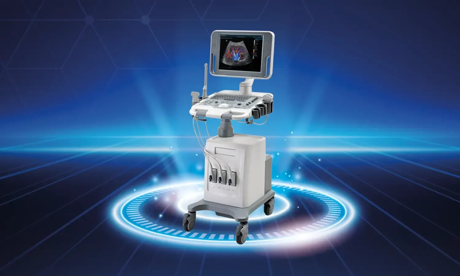
4D ultrasound is a method of obtaining real-time, moving three-dimensional images of the baby by processing sound waves at high speed. Unlike a frozen image from a 3D ultrasound, time is added as a dimension in 4D devices; thus, the baby's yawning, thumb-sucking, facial expressions, and movements can be observed live.
Working Principle
High-frequency sound waves sent from the ultrasound device reflect off tissues in the womb at different densities. The computer processes these reflections with mathematical algorithms, converts them into volumetric data, and transfers them to the screen in real-time. Since the procedure is based entirely on sound waves, it does not contain radiation and can be safely applied throughout pregnancy.
In Which Situations Is It Used?
4D ultrasound offers valuable medical information beyond the purpose of simply showing the baby to the expectant mother. Its main areas of use are as follows:
- Detailed evaluation of the baby's face, lip, and palate structure and early detection of anomalies such as cleft lip and palate
- Structural examination of extremities such as hands, feet, and fingers
- Detailed imaging of heart, brain, and spine development
- Observation of movement patterns that are important for the baby's neurological development
- Evaluation of the placenta, umbilical cord, and amniotic fluid
- Separate examination of babies in multiple pregnancies
Advantages of 4D Ultrasound
4D imaging provides significant advantages in terms of both medical diagnosis and establishing family bonding:
- High image quality allows for earlier and clearer detection of structural anomalies
- Evaluation of the baby's facial expressions and behaviors with moving images
- Enabling the expectant mother and father to form a stronger emotional bond with their baby
- Capturing some details that might be missed in a classic ultrasound
- Providing the baby's images to families as a video
- Being completely safe for the mother and baby as it is a non-invasive procedure
Ideal Application Time and Process
4D ultrasound can be performed at any stage of pregnancy; however, the period when the baby's facial features and organs are imaged most clearly is usually between 26 and 32 weeks of gestation. During these weeks, the baby's subcutaneous adipose tissue is sufficiently developed and the amount of amniotic fluid is at an appropriate level, so the images are obtained in great detail.
Procedure Process
The application is performed similarly to a classic ultrasound examination. The expectant mother is placed in a supine position, a special gel is applied to the abdominal area, and imaging is performed with the help of a probe. The procedure takes about 20-30 minutes and is painless and comfortable. If the baby's position makes imaging difficult, the expectant mother may be asked to move a little or wait for a short time.
Points to Consider
To get the most efficient results from a 4D ultrasound, it is recommended to pay attention to some points:
- Not being excessively full or hungry before the procedure is important for image quality
- Consuming plenty of water supports obtaining clearer images by increasing amniotic fluid
- If the baby's position is not suitable, the session can be repeated; this is not a cause for concern
- 4D ultrasound does not replace routine pregnancy follow-up and detailed second-level ultrasound scans; it is complementary to them
- It is not recommended to be applied very frequently unless medically necessary
Differences Between 4D Ultrasound and Classic Ultrasound
While classic 2D ultrasound provides cross-sectional black-and-white images of tissues, 3D devices make these cross-sections volumetric, and 4D devices add the time dimension to create live, moving images. 2D ultrasound continues to be the gold standard for many measurements and structural evaluations; 4D imaging does not replace it, it is a complementary tool.
In 4D imaging, surface structures such as the face, hands, and feet can be observed in a much more natural way. However, the detail of 2D images is still indispensable for the evaluation of internal organs, flow measurements, and structural scans. The ideal approach is to use both methods together in pregnancy follow-up.
Frequently Asked Questions for Expectant Mothers
- Does it harm the baby? No; since sound waves are used, it does not contain ionizing radiation and is safe when applied for an appropriate duration.
- Should it be done in every pregnancy? It is not mandatory unless there is a medical necessity; however, it provides valuable information in pregnancies with risk factors.
- Is the baby's gender clearly visible? It can usually be viewed with high accuracy from the 16-20th week onwards, provided the baby is in a suitable position.
- Are the results given immediately? Evaluation is made during the procedure, and the detailed report and images are delivered shortly after.
The Future of 4D Ultrasound and Technological Developments
Imaging technologies are advancing every day; HD live ultrasound, real-time 5D imaging, and artificial intelligence-supported analysis systems have begun to enter clinical use. While these new-generation devices present the baby's facial features with the clarity of a photograph, they facilitate the physician's work in heart, brain, and other organ evaluations thanks to automatic measurement algorithms.
Artificial intelligence-supported image analysis also plays an important role in detecting subtle anomalies that are difficult to notice on ultrasound. In the coming years, the accuracy and reliability of pregnancy follow-up will increase even further as these technologies become more widespread. Choosing the right center and an experienced physician is of great importance for expectant mothers to get the highest benefit from this process.
4D Ultrasound at Koru Hospital
As Koru Hospital, we bring together state-of-the-art imaging devices and our experienced obstetricians and gynecologists in pregnancy follow-up. In our 4D ultrasound applications, we both evaluate the baby's health status in detail and provide our expectant mothers with unforgettable moments with their babies. We are with you at every stage of your pregnancy with our modern device infrastructure, expert staff, and patient-oriented approach.