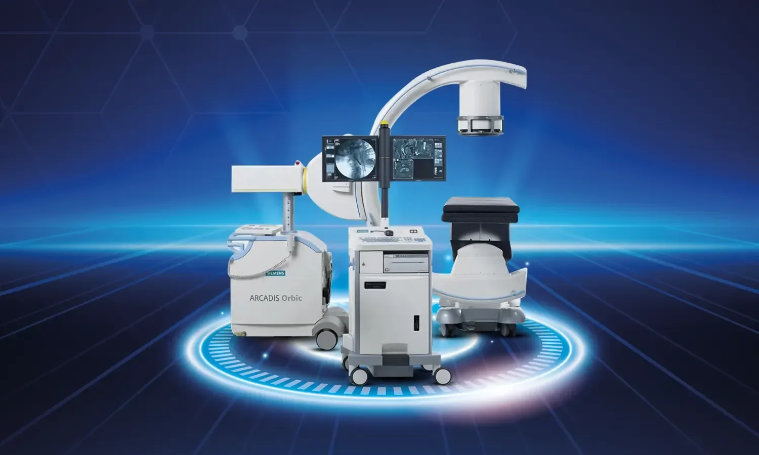
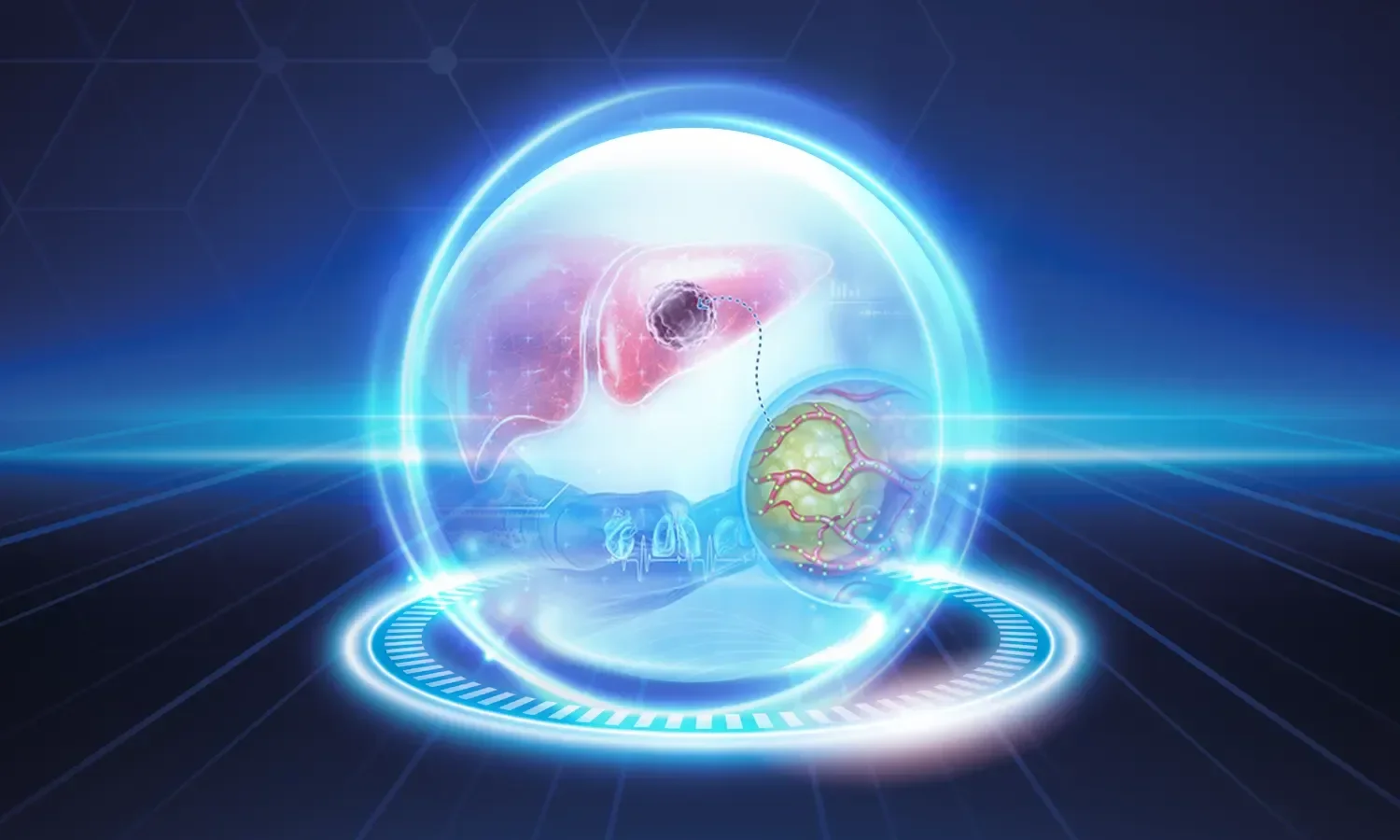
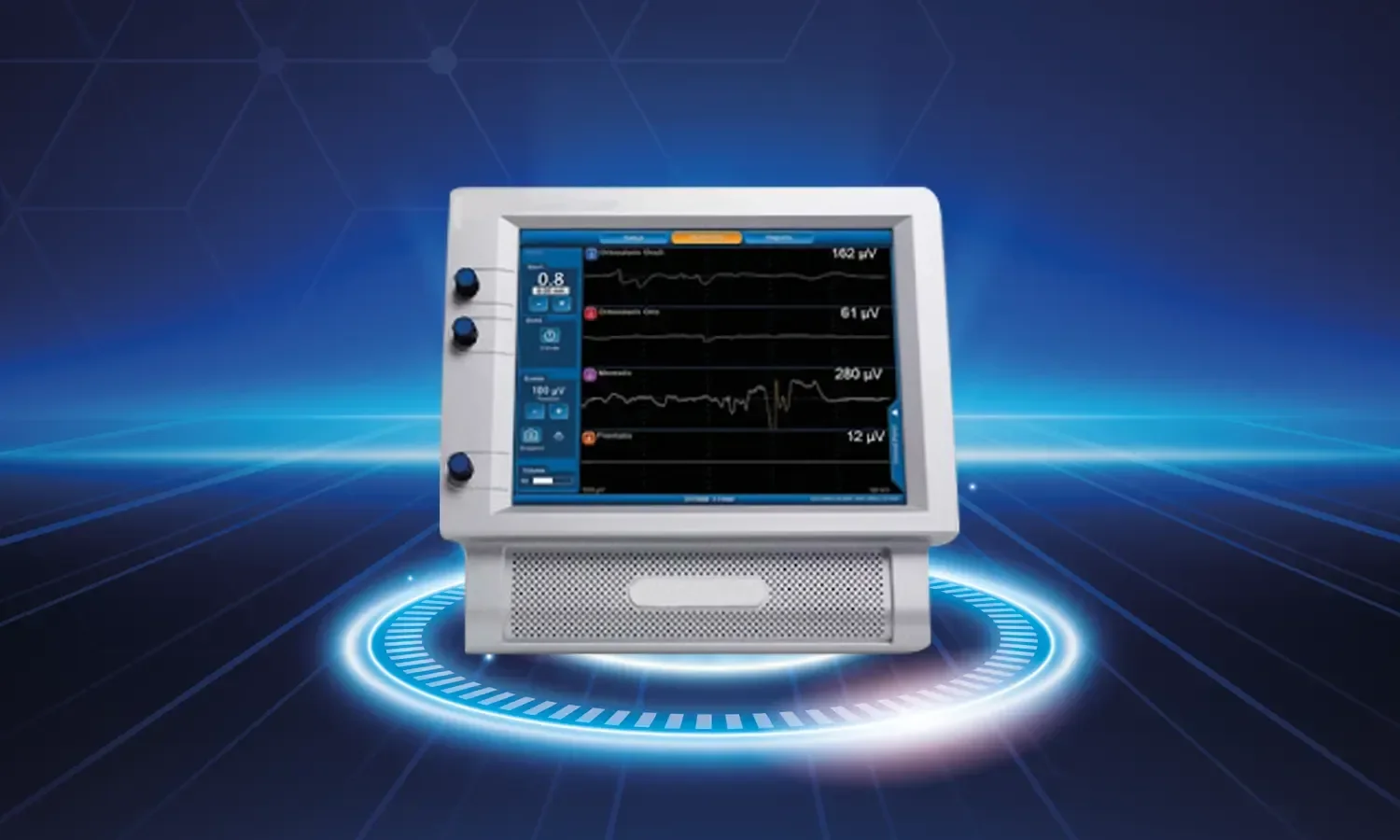
Neuronavigation is a three-dimensional imaging and real-time guidance technology used in brain and spinal surgery. Working similarly to a GPS system, this advanced technology enables surgeons to move with sub-millimeter precision, serving as a vital guide in complex operations such as brain tumors, aneurysms, epilepsy surgery, and spinal surgery. Having become an integral part of modern neurosurgery, this system offers patients safer and less invasive treatment options. By enabling safe access even to deep brain lesions that are difficult to reach with traditional surgical methods, neuronavigation has significantly increased neurosurgical success rates in recent years.
What is Neuronavigation and How Does It Work?
Neuronavigation systems convert magnetic resonance (MR) and computed tomography (CT) images obtained from the patient before surgery into a three-dimensional model using computer software. This model creates a virtual map of the patient's brain or spine anatomy. During surgery, the position of the instruments used by the surgeon is continuously tracked via special infrared cameras and reference markers. The system displays the location of the surgical instrument tips on the screen in real-time, allowing the surgeon to clearly see the location of critical surrounding anatomical structures while reaching the target area.
Working Principle of the System
The workflow consists of three main stages:
- Imaging stage: High-resolution MR and CT images are acquired; functional MR (fMRI), tractography (DTI), and PET data are added when necessary.
- Planning stage: The surgeon determines the safest entry path, the target area, and the location of critical structures on the virtual model.
- Registration stage: The alignment between the patient's actual anatomy and the virtual model is established in the operating room.
- Navigation stage: During surgery, the real-time position of surgical instruments is displayed instantly on the three-dimensional model on the screen.
- Verification: The accuracy of the system is checked at critical points using intraoperative imaging.
In Which Diseases Is It Used?
Neuronavigation technology is successfully applied in a wide range of diseases:
- Brain tumors: Gliomas, meningiomas, metastatic tumors, and pituitary adenomas.
- Vascular pathologies: Brain aneurysms and arteriovenous malformations (AVM).
- Epilepsy surgery: Detection and removal of the seizure focus.
- Spine surgery: Pedicle screw placement and spinal tumor resection.
- Movement disorders: Placement of deep brain stimulation (DBS) electrodes for Parkinson's disease.
- Hydrocephalus: Shunt catheter placement and endoscopic third ventriculostomy.
- Biopsy procedures: Sampling from deep-seated lesions.
- Skull base surgery: Safe access to complex anatomical regions.
- Craniofacial surgery: Facial skeleton reconstruction.
- Pediatric neurosurgery: Conditions requiring precise surgery in pediatric patients.
Advantages of Neuronavigation
The clinical benefits provided by this advanced technology are extremely comprehensive:
- High precision: Sub-millimeter accuracy ensures the protection of critical structures.
- Smaller surgical incision: Tissue damage is reduced thanks to a minimally invasive approach.
- Reduced risk of complications: Blood vessels, nerves, and functional brain areas are preserved.
- Efficiency in tumor removal: Clearly defining the tumor boundaries increases the rate of total resection.
- Faster recovery process: Patients are discharged sooner due to shorter operation times and reduced trauma.
- Increased surgical safety: The impact of unexpected anatomical conditions is minimized thanks to a pre-planned route.
- Preservation of eloquent (speech, movement) areas: Avoiding damage to functional brain areas.
- Reduced surgical time: Effective planning shortens the operation duration.
- Training and simulation opportunity: It is a valuable learning tool for young surgeons.
Treatment Process
During the surgery performed with neuronavigation, the patient first undergoes detailed imaging examinations. High-resolution MR, CT, functional MR, and, if necessary, tractography examinations are completed. The obtained data is uploaded to the neuronavigation system, and detailed planning is carried out by the surgical team. In this planning, the safest entry path, the strategy to reach the target area, and the critical structures that must be protected are determined. After the patient is fixed in the appropriate position in the operating room, the reference frame is placed, and the system is calibrated. The patient's actual anatomy is matched with the virtual model (registration process). Throughout the operation, the surgeon tracks the position of the instruments used on the three-dimensional model on the screen in real-time. The system is updated with intraoperative MR or ultrasound when necessary. The success of the treatment is evaluated with post-operative control imaging, and the patient is followed up in the intensive care unit.
Points to Consider
Although neuronavigation is a highly reliable technology, some important points should be kept in mind. The fact that brain tissue can change position during surgery (brain shift) may affect the accuracy of the system. For this reason, experienced teams continuously check accuracy with additional methods such as intraoperative MR or ultrasound. Maintaining patient immobility, correct calibration, and the currency of the images are critical factors determining success. The system does not replace the surgeon's experience; anatomical knowledge and surgical judgment are always paramount. It is important that pre-operative images are sufficiently recent and that the patient's conditions that create MR incompatibility, such as pregnancy or a pacemaker, are evaluated.
Differences Between Neuronavigation and Classical Surgery
While the surgeon acts using anatomical knowledge and visual references in classical brain and spine surgery, surgery with neuronavigation provides real-time spatial information. This difference is of great importance, especially in lesions that are deep-seated, small, or intertwined with critical structures. In classical surgery without neuronavigation, even an experienced surgeon may lose anatomical landmarks in some cases; however, this risk is minimized with neuronavigation. Modern systems also allow for the mapping of the brain's speech, movement, and memory areas by combining them with diffusion and functional imaging. When the awake craniotomy technique is combined with neuronavigation in tumors passing through eloquent cortical areas, the patient's functions can be tested even during the operation. This combination is revolutionary, especially in gliomas in the dominant hemisphere. The learning curve of surgeons is also significantly shortened thanks to neuronavigation, and it is becoming standard in training centers.
As Koru Hospital, we provide services at the highest standards in the field of brain and spine surgery with state-of-the-art neuronavigation systems in our neurosurgery department. With our experienced neurosurgeons, multidisciplinary team, and modern operating room infrastructure, we provide our patients with safe, effective, and minimally invasive treatment options. We meticulously apply advanced imaging and navigation technologies to meet our patients' expectations at the highest level and maximize surgical success.