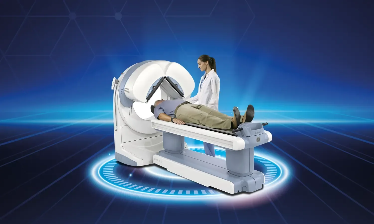
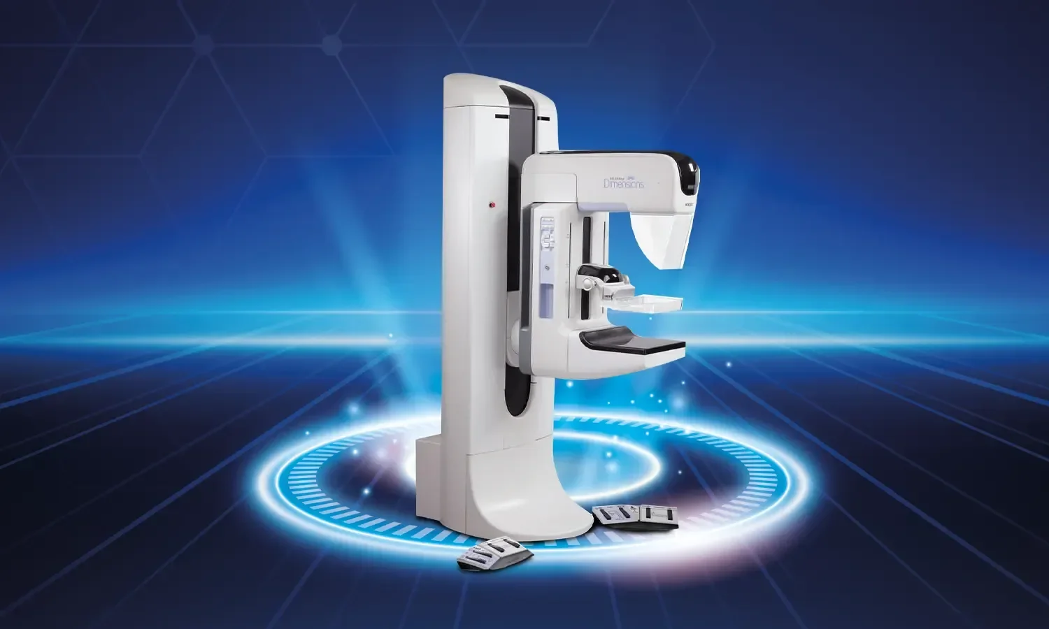
Micro-ultrasound fusion biopsy is an advanced imaging and sampling method used in the diagnosis of prostate cancer. Traditionally, random tissue sampling (biopsy) performed under transrectal ultrasound (TRUS) guidance is used to diagnose prostate cancer. However, this method can sometimes miss cancerous areas or lead to sampling from clinically insignificant regions. Fusion biopsy has been developed to overcome these limitations and has become the standard in modern urological practice.
How Does Micro-Ultrasound Fusion Biopsy Work?
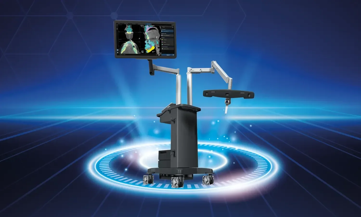
In this technique, detailed prostate images obtained via multiparametric magnetic resonance imaging (mpMRI) are fused with real-time ultrasound images. This allows for a clearer view of the prostate's structure and potential cancerous areas. Micro-ultrasound technology operates at a much higher frequency than traditional ultrasound, enabling the visualization of the prostate's internal structure with micron-level detail, thus allowing suspicious areas to be detected directly.
The system enables the computer-assisted overlay of suspicious areas marked on the MRI onto the ultrasound image, facilitating targeted biopsy. As a result, small but clinically significant cancers that could not be detected by random sampling in previous years can now be accurately diagnosed.
Procedure Steps
- Magnetic Resonance Imaging (MRI): The first step is to obtain a detailed multiparametric magnetic resonance image of the prostate. These images are used to identify potential cancerous areas.
- Ultrasound Imaging: Subsequently, live images of the prostate are obtained using real-time ultrasound.
- Image Fusion: MRI images are fused with ultrasound images. This helps the physician see the internal structure of the prostate more clearly and identify potential cancerous regions.
- Targeted Biopsy: Tissue samples are taken from the identified potential cancerous areas. These samples are examined in the laboratory to determine the type and aggressiveness of the cancer, if present.
- Systematic Sampling: In addition to the targeted biopsy, systematic samples are also taken from the remaining prostate tissue to increase safety.
Which Patients Is It Recommended For?
- Patients with high PSA levels but negative standard biopsy results
- Cases where suspicion of prostate cancer persists despite a normal previous biopsy
- Patients with suspicious lesions (PI-RADS 3, 4, or 5) detected on mpMRI
- Low-risk prostate cancer patients under active surveillance
- Men in the high-risk group with a family history of prostate cancer
- Patients with suspected recurrence after prostate cancer treatment
- Individuals carrying genetic risk factors such as BRCA mutations
- Patients who have previously undergone prostate surgery and are being evaluated for recurrence
Advantages of Micro-Ultrasound Fusion Biopsy
- More precise targeting: Detailed prostate images obtained via mpMRI are fused with real-time ultrasound images, allowing cancerous areas to be targeted more accurately.
- Higher diagnostic sensitivity: The detection rate of clinically significant cancers increases; unnecessary biopsies are reduced.
- Reduced complications: Since less tissue may need to be removed due to more precise targeting, the risk of bleeding, infection, and other complications is reduced.
- Fewer unnecessary biopsies: mpMRI scans more clearly identify potential cancerous regions, reducing the collection of unnecessary samples.
- Treatment planning and monitoring: Used not only for cancer diagnosis but also for staging and post-treatment follow-up.
- Patient satisfaction: Offers a less invasive procedure, lower risk of complications, and less discomfort.
- Reduced detection of clinically insignificant cancers: Prevents overtreatment.
- Accurate follow-up opportunity for active surveillance patients
The Procedure Process
Before the fusion biopsy, the patient undergoes a multiparametric prostate MRI, and the images are evaluated using specialized software. Suspicious areas (using PI-RADS scoring) are marked. The procedure is usually performed under local anesthesia or light sedation; a micro-ultrasound probe is inserted via the transrectal or transperineal route. The transperineal approach is a modern technique that can further reduce the risk of infection. MRI images are registered with ultrasound images in real-time, and samples are taken from the target areas using a fine needle. Both targeted and systematic sampling can be performed in the same session.
The procedure typically takes 20-40 minutes. Afterward, the patient is sent home following a short observation period. Pathology results are usually ready within a few business days. Once the results are evaluated, a treatment plan is created by a multidisciplinary team consisting of urology, radiation oncology, and medical oncology specialists. Depending on the stage and aggressiveness of the cancer and the patient's general condition, treatment options can be planned as active surveillance, surgery, radiotherapy, or combined therapies.
Points to Consider
Although fusion biopsy is a minimally invasive and safe procedure, as with any biopsy procedure, rare cases may involve mild bleeding, blood in the urine, infection, or temporary urinary retention. Blood-thinning medications used before the procedure must be reported to the physician and discontinued for an appropriate period if necessary. Antibiotic prophylaxis reduces the risk of infection. Pathology results are usually ready within a few days, and the treatment plan is determined based on these results. It is normal and temporary to see blood in the ejaculate for a few weeks after the procedure. Patients are advised to consume plenty of fluids after the procedure, complete the antibiotic treatment recommended by the physician, and contact the hospital if there is a change in their symptoms.
Fusion Biopsy at Koru Hospital
As Koru Hospital, we perform micro-ultrasound fusion biopsy at high standards with our experienced specialist staff in the fields of urology and radiology. With our modern mpMRI system, advanced fusion software, and reliable pathology laboratory, we aim to achieve accurate and fast results in the diagnosis of prostate cancer. Our patients are followed by our multidisciplinary team throughout the entire process, from pre-procedure to pathology results; treatment planning is carried out through the joint evaluation of urology, radiology, radiation oncology, medical oncology, and pathology specialists. Each patient's individual clinical condition is discussed in detail in our tumor board, and a personalized treatment plan is created in line with the most current evidence-based guidelines. We continue to stand by our patients with our comfortable, patient-centered approach, experienced healthcare personnel, and continuous psychological support opportunities. As Koru Hospital, our goal is to ensure the treatment of prostate cancer with the right diagnosis at the right stage and to maintain our patients' quality of life at the highest level.